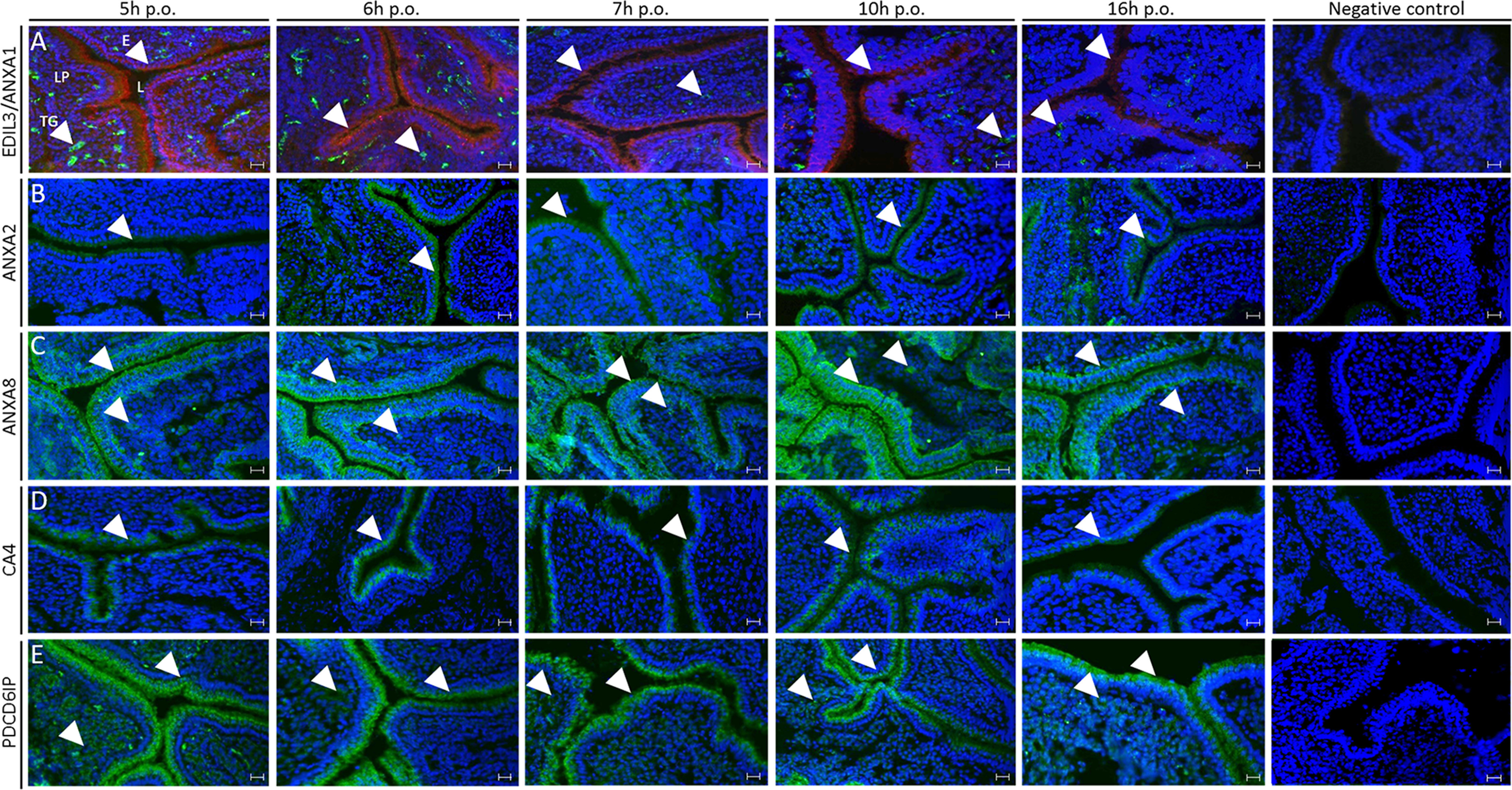
Figure 6.

Immunofluorescence of ANXA1, ANXA2, ANXA8, EDIL3, CA4, and PDCD6IP in chicken uterus. The respective rows correspond to: A, co-staining of ANXA1 (red) and EDIL3 (green), B, staining of ANXA2 (green); C, staining of ANXA8 (green); D, staining of CA4 (green); and E, staining of PDCD6IP (green). ANXA1, ANXA2, and CA4 were localized in the epithelium. ANXA8 and PDCD6IP signals were observed in both epithelium and tubular glands of lamina propria. EDIL3 was solely detected in tubular glands. E, ciliated and glandular epithelium; L, lumen; LP, lamina propria; TG, tubular glands. The primary and secondary antibodies are compiled in Table S4. Bars, 100 μm. White arrowheads indicate positive signal in either tubular glands or epithelium.
