
FIGURE 2.
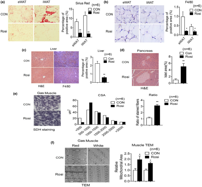
Rosiglitazone treatment in aging mice reduces inflammation, increases oxidative fiber number, and mitochondrial area in muscle. (a–i) Analysis of 20‐month‐old mice after 6 months of treatment, either with control diet or diet supplemented with Rosi (1 mg/kg of mouse weight/day) (n = 6). Representative image of Sirius Red staining (a) and F4/80 staining (b) of eWAT and iWAT with quantification. (c) Representative H&E images and F4/80 staining of liver with quantification. (d) Representative H&E images of pancreas and quantification of islet area. (e) Cross‐sectional area (CSA) of fiber of gastrocnemius (Gas) muscle by Succinic dehydrogenase (SDH) staining and ratio of oxidative fibers. (f) Representative images of gastrocnemius muscle by transmission electron microscopy and quantification of mitochondrial area. Data are presented as mean ± SEM and Student t tests were performed to compare control and Rosi‐treated group. (*p < 0.05, **p < 0.01)