Figure 2.

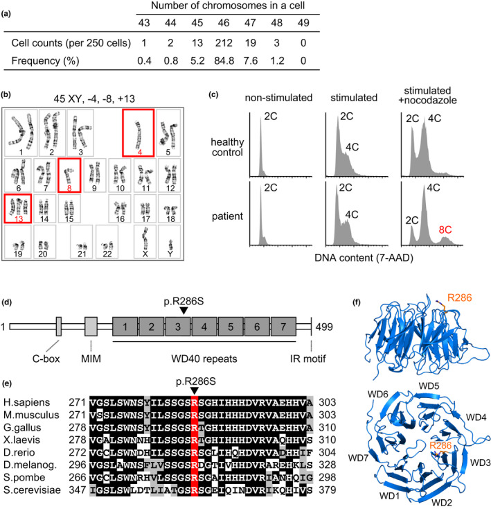
Chromosome number instability and CDC20 mutation in the patient's peripheral blood mononuclear cells. (a) Chromosome number instability observed in the patient's peripheral blood mononuclear cells (PBMCs). (b) A representative karyotype of the patient's PBMCs. Red squares indicate chromosome gain or loss. (c) Flow‐cytometric analyses of DNA contents in the PBMCs of the patient and a healthy control with or without mitotic stimulation and nocodazole treatment. X‐axes indicate nuclear DNA contents (2C, diploid; 4C, tetraploid; 8C, octoploid) stained with 7‐amino‐actinomycin D (7‐AAD) on a linear scale. (d) A schematic representation of the human CDC20 protein. The R286 amino acid residue is indicated with a black arrowhead. MIM, MAD2 interacting motif. (e) A cross‐species amino acid sequence comparison of CDC20 around the R286 residue (red). Identical and conserved amino acids are marked with black and gray boxes, respectively. (f) Side and top view of the WD40 repeat propeller domains (WD1–7) of the human CDC20 protein. The R286 residue (orange stick) is located at the top of WD3.
