Figure 5.

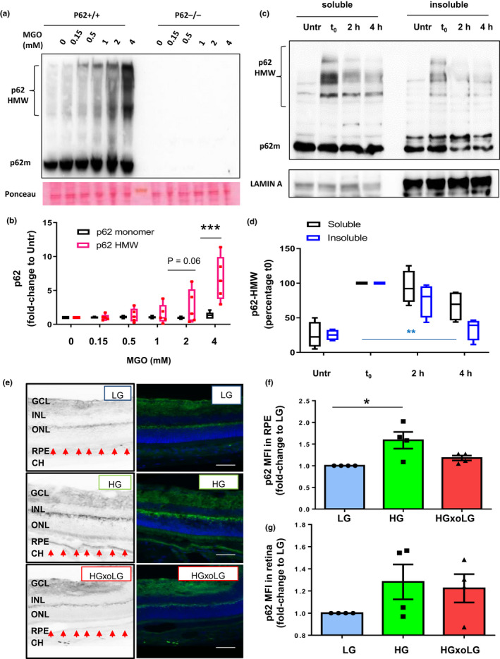
Accumulation of high molecular weight p62 upon glycative stress is reversible. (a,b) WT MEFs (p62+/+) and MEFs lacking p62 (p62−/−) were incubated with the indicated concentration of MGO for 2 hours, and whole cellular extracts were immunoblotted against p62. (a) Representative immunoblot and (b) quantification of p62 monomer and high molecular weight p62 (HMW‐p62) values relative to untreated cells are shown. Values are mean ±SEM (n = 5). We observed an interaction (p < 0.0001) between the MGO concentration and the HMW‐p62 using two‐way ANOVA analysis. The differences between HMW‐p62 and monomeric p62 after the Sidak's multiple comparison test were significant for the 4 mM doses of MGO (***p < 0.001). (c,d) ARPE‐19 cells were treated with 2 mM MGO for 2 hours followed by incubation in complete medium (no MGO) for either 2 or 4 hours. Cellular lysates were subjected to extraction with 1% Triton X‐100 and soluble and insoluble fractions were immunoblotted for the indicated proteins. (c) Representative immunoblot and (d) quantification of HMW‐p62 values relative to untreated cells are shown. Values are mean ±SEM (n = 5). Differences between t0 and insoluble p62 were significant for the 4 mM doses of MGO using one‐way ANOVA followed by Dunnett's multiple comparison test (**p < 0.01). (e,f) Retinal tissue sections from low‐glycemic (LG), high glycemic (HG), and crossover diet (HGxoLG) were analyzed immunohistochemically for p62. (e) Representative images of p62 immunostaining and mean intensity fluorescence in (f) the retinal pigment epithelial layer and (g) neuroretina relative to values in LG‐diet are shown. Values are mean ±SEM (n = 4). Abbreviations: CH, choroid; RPE, retinal pigment epithelium; INL, inner nuclear layer; IPL, inner plexiform layer; ONL, outer nuclear layer; GCL, ganglion cell layer. p < 0.05 in one‐way ANOVA followed by Dunnett's multiple comparison test
