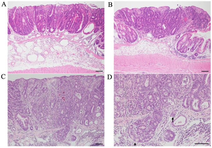
Figure 2.
Histopathology of colon tumors from AOM/DSS mice at weeks 10, 20 and 30. Colon tumors of AOM/DSS mice had the typical appearance of adenocarcinoma. Compared with AOM/DSS mice at weeks (A) 10 and (B) 20, (C and D) tumor infiltrations into the submucosal layer (arrowhead) and tumor invasions into vessels (arrow) were more frequently observed in AOM/DSS mice at week 30. Histopathology was performed using hematoxylin and eosin staining. Original magnification, (A-C) ×100 and (D) ×200. Scale bars, 100 µm. AOM, azoxymethane; DSS, dextran sodium sulfate.

