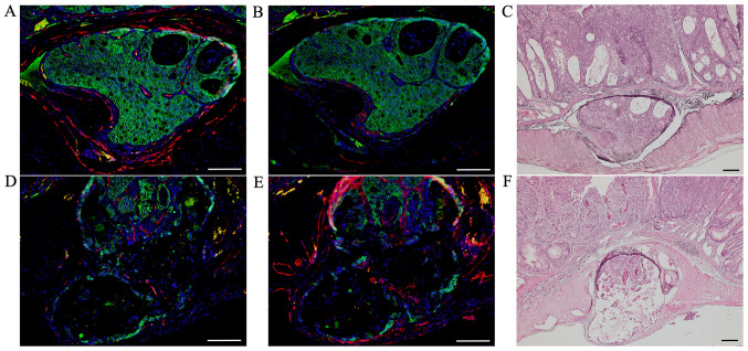
Figure 3.
Double immunofluorescent staining of β-catenin (green) with CD34 (red) and podoplanin (red) in AOM/DSS mice at week 30. DAPI (blue) was used for nuclear staining. (A-C) In AOM/DSS mice that showed blood vessel invasion, (A and B) β-catenin-positive cells were diffusely distributed throughout the tumors in vessels. (A) Although immunofluorescent staining of CD34 revealed ring-shaped positivity around the vessel lumens, (B) podoplanin-positive cells were not detected in these vessels. (D-F) In AOM/DSS mice that showed lymph vessel invasion, (D and E) β-catenin-positive tumor cells were similarly distributed in vessels. (D) Contrarily, CD34-positive cells were not detected in the vessels, and (E) immunofluorescent staining of podoplanin demonstrated ring-shaped positivity around the vessel lumens. (C and F) Following immunofluorescent staining, the same sections were stained with hematoxylin and eosin, and observed for tumor invasion into vessels by light microscopy. Original magnification, (A, B, D and E) ×200 and (C and F) ×100. Scale bars, 100 µm. AOM, azoxymethane; DSS, dextran sodium sulfate.

