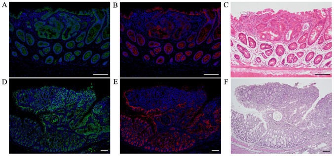
Figure 4.
Double immunofluorescent staining for β-catenin (green) and E-cadherin (red) in AOM/DSS mice at weeks 10 and 30. DAPI (blue) was used for nuclear staining. Immunofluorescent staining of β-catenin revealed weak positivity only in the cell membrane of non-tumorous mucosae in AOM/DSS mice at weeks (A) 10 and (D) 30. Strongly β-catenin-positive cells were distributed throughout the tumors in AOM/DSS mice, and their expression was predominantly observed in the cytoplasm and nucleus of tumor cells. In the same sections, immunofluorescent staining of E-cadherin demonstrated strong positivity in the cell membrane of non-tumorous mucosae in AOM/DSS mice at weeks (B) 10 and (E) 30. Positive levels of E-cadherin in the cell membrane of colon tumors in AOM/DSS mice were clearly reduced as compared with those of non-tumorous mucosae. (C and F) After immunofluorescent staining, the same sections were stained with hematoxylin and eosin, and colon tumors were confirmed using light microscopy. Original magnification, (A-C) ×200 and (D-F) ×100. Scale bars, 100 µm. AOM, azoxymethane; DSS, dextran sodium sulfate.

