Figure 5.
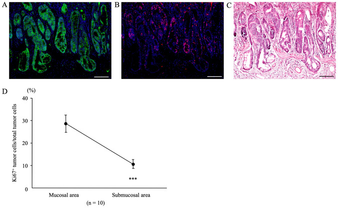
Double immunofluorescent staining for β-catenin (green) and Ki67 (red) in sites of submucosal tumor infiltration of AOM/DSS mice at week 30. DAPI (blue) was used for nuclear staining. (A) β-catenin-positive tumor cells were diffusely distributed in both mucosal areas and sites of submucosal infiltration in AOM/DSS mice at week 30. (B) For the same sections, immunofluorescent staining of Ki67 was performed. (C) Following immunofluorescent staining, the same sections were stained with hematoxylin and eosin, and colon tumors and muscularis mucosae were confirmed under a light microscope. Original magnification, ×200. Scale bars, 100 µm. (D) Ki67-positive tumor cells/total tumor cells in mucosae and submucosae. The percentage of Ki67-positive tumor cells in mucosal areas of AOM/DSS mice (28.66±3.80%) was significantly higher than that in sites of submucosal infiltration (10.66±1.97%; n=10; P=0.0001). Data are presented as the mean ± standard error of the mean, and were analyzed using a paired t-test. ***P<0.001. AOM, azoxymethane; DSS, dextran sodium sulfate.

