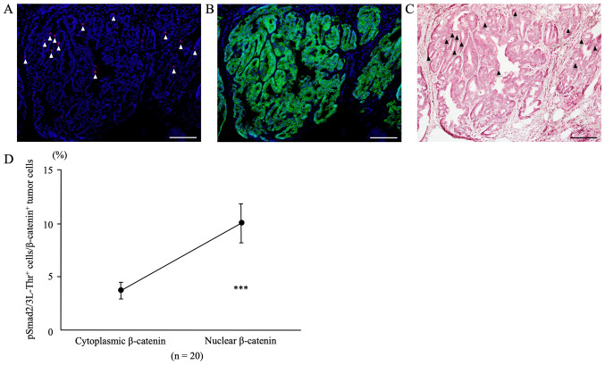
Figure 7.
Double immunofluorescent staining for pSmad2/3L-Thr (red; arrowheads) and β-catenin (green) in colon tumors from azoxymethane/dextran sodium sulfate mice at week 30. DAPI (blue) was used for nuclear staining. (A) pSmad2/3L-Thr-positive cells (arrowhead) were scattered among tumor cells. (B) For the same sections, immunofluorescent staining of β-catenin revealed cytoplasmic and nuclear β-catenin expression in tumor cells. (C) Following immunofluorescent staining, the same sections were stained with hematoxylin and eosin, and colon tumors and pSmad2/3L-Thr-positive cells (arrowheads) were confirmed by light microscopy. Original magnification, ×200. Scale bars, 100 µm. (D) pSmad2/3L-Thr-positive cells/β-catenin-positive tumor cells within cytoplasm and nuclei. The percentage of pSmad2/3L-Thr-positive cells among the nuclear β-catenin-positive tumor cells (9.98±1.82%) was significantly higher than that among the cytoplasmic β-catenin-positive tumor cells (3.67±0.77%; n=20; P=0.0001). Data are presented as the mean ± standard error of the mean, and were analyzed using a paired t-test. ***P<0.001.

