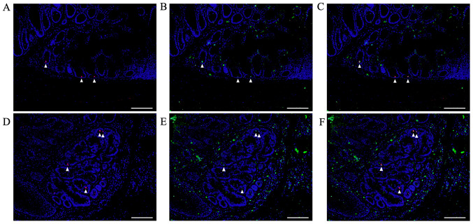
Figure 8.
Double immunofluorescent staining for pSmad2/3L-Thr (red; arrowheads) and Bmi1 (green) in non-tumorous mucosae and CRCs of AOM/DSS mice at week 30. DAPI (blue) was used for nuclear staining. (A) pSmad2/3L-Thr-positive cells were sparsely detected around crypt bases in non-tumorous mucosae. (D) In colon tumors from AOM/DSS mice, pSmad2/3L-Thr-positive cells were scattered among tumor cells. pSmad2/3L-Thr-positive cells exhibited immunohistochemical co-localization with Bmi1 in both (B and C) non-neoplastic and (E and F) neoplastic epithelial cells, (C and F) as indicated in the merged panels. Original magnification, ×200. Scale bars, 100 µm. AOM, azoxymethane; DSS, dextran sodium sulfate.

