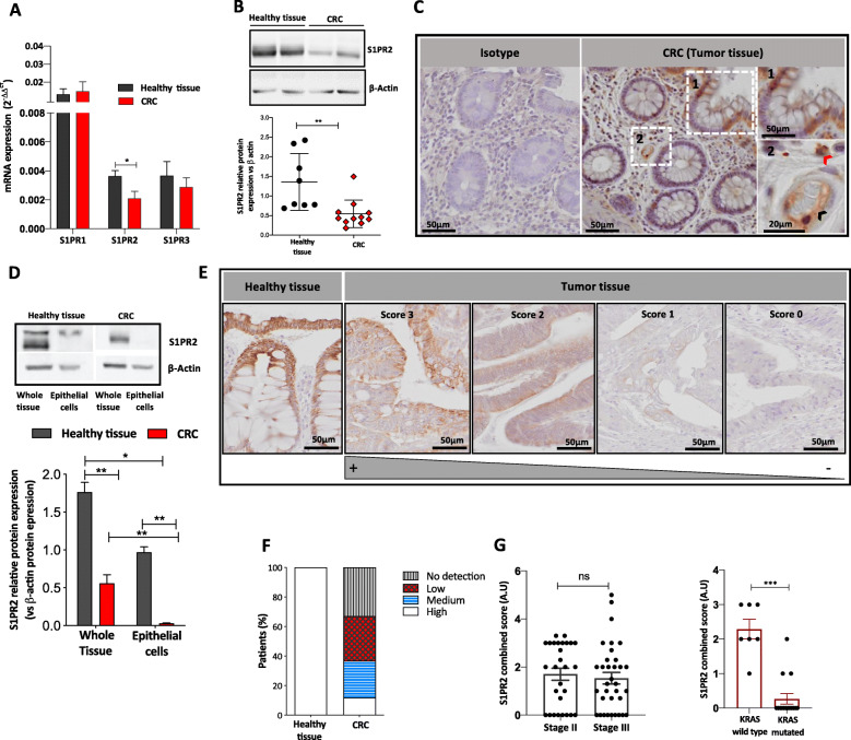
Fig. 1.
S1PR2 expression in human colorectal cancer. a Relative mRNA expression levels of S1PR1, S1PR2 and S1PR3 in CRC (n = 39) with stage II/III pT1-T4, and normal colon tissue (n = 16) samples. mRNA data are presented as the mean ± SEM and normalized to the expression of human GAPDH and expressed as 2-ΔΔCt. The significance was evaluated by two-way ANOVA followed by Bonferroni’s test; *p < 0.05. b Representative examples of Western blot analysis (up panel) and densitometric analysis (low panel) of S1PR2 in human CRC (n = 11) and normal colonic mucosa (n = 8) samples. Protein levels of S1PR2 were normalized on the β-actin expression. Mean ± SEM, **p = 0.002 by Mann-Whitney test. c Representative images of S1PR2 immunostaining of healthy colonic mucosa. In the left panel is reported the isotype, whereas in the right panel is evidenced the positive staining for S1PR2 in the epithelium, endothelial cells (black arrow), and immune cells (red arrow). d Analysis of S1PR2 in whole tissue and primary epithelial cells isolated from adenocarcinomas and adjacent healthy tissue (n = 8). Protein levels of S1PR2 were normalized on the β-actin expression. e Immunohistochemical analysis of S1PR2 expression in CRC samples was assessed using a combined score between intensity and extension of the immunoreaction. The images were acquired by the DotSlide system at 20x objective. f The stacked bar chart reports the percentage of cases expressing the different intensity of immunostaining in normal (Healthy tissue) and CRC samples. S1PR2 combined score in correlation to (f) tumor stage II/III and (g) KRAS mutation in CRC. Means ± SEM, *p < 0.05, **p < 0.01, and ***p < 0.001 by one-way ANOVA followed by Bonferroni’s test

