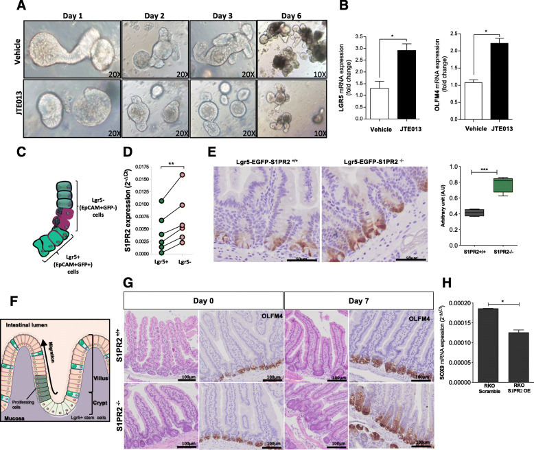
Fig. 5.
Deletion of S1PR2 promotes the expansion of intestinal stem cells. a Organoid development in the presence of JTE013 (10 μM) or vehicle over 6 days. Images were acquired by an inverted light microscope at 10 and 20x objectives. b mRNA analysis of LGR5 and OFLM4 in organoids on day 6. c Schematic distribution of epithelial progenitor stem cells Lgr5+ (EPCAM+GFP+) and differentiated epithelial cells Lgr5- (EPCAM+GFP-) in the crypts of Lgr5-EGFP-IRES-creERT2 mice. d S1PR2 mRNA expression in sorted EPCAM+ GFP positive (Lgr5+) and EPCAM+ GFP negative (Lgr5-) isolated from Lgr5-EGFP-IRES-creERT2 mice (n = 6). Data as a mean ± SEM of 6 independent experiments. ** p = 0.003 by a paired t-test. e Immunostaining of LGR5 and its quantification on intestinal sections of Lgr5-EGFP-S1PR2−/− and Lgr5-EGFP-S1PR2+/+ mice. f Schematic overview of intestinal epithelial regeneration showing expansion and differentiation of Lgr5 stem cells that move upward into the villus allowing a rapid regeneration of the epithelium. g Impaired expression of OLFM4 in the small intestine of S1PR2−/− and S1PR2+/+ mice at day 0 and 7 of X-ray irradiation. Data are mean ± SEM and representative of 3 experiments (n = 4). h mRNA levels of SOX9 in RKO-OE and scramble cells, presented as mean ± SEM, normalized to the expression of GAPDH and expressed as 2-ΔΔCt. *p < 0.05; ** p < 0.01; and ***p < 0.001 by Mann-Witney test

