Fig. 4.
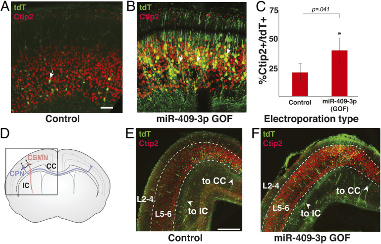
miR-409-3p promotes CSMN subtype identity and subcerebral axon trajectory in vivo. Representative fluorescence micrographs of e18.5 cortices electroporated at e13.5 illustrate an increase in the percent layer V CTIP2+/tdT+ neurons (CSMN, arrows) with miR-409-3p GOF (B), compared to scrambled control (A). (Scale bar, 50 μm.) (C) miR-409-3p GOF results in a 100% increase (43.3% from 21.5%) in CTIP2+/tdT+ neurons (CSMN), compared to scrambled control in vivo. Error bars represent SEM. (D) Schematic of layer V CSMN (red) projecting subcerebrally via the internal capsule (IC) and CPN (blue) projecting interhemispherically via the corpus callosum (CC). (E and F) Representative coronal fluorescence micrographs of e18.5 brains electroporated at e13.5 illustrate many more axons projecting subcerebrally via the IC and very few apparent axons projecting interhemispherically via the CC in miR-409-3 GOF (F) compared to in scrambled control (E). (Scale bar, 500 μm.)

