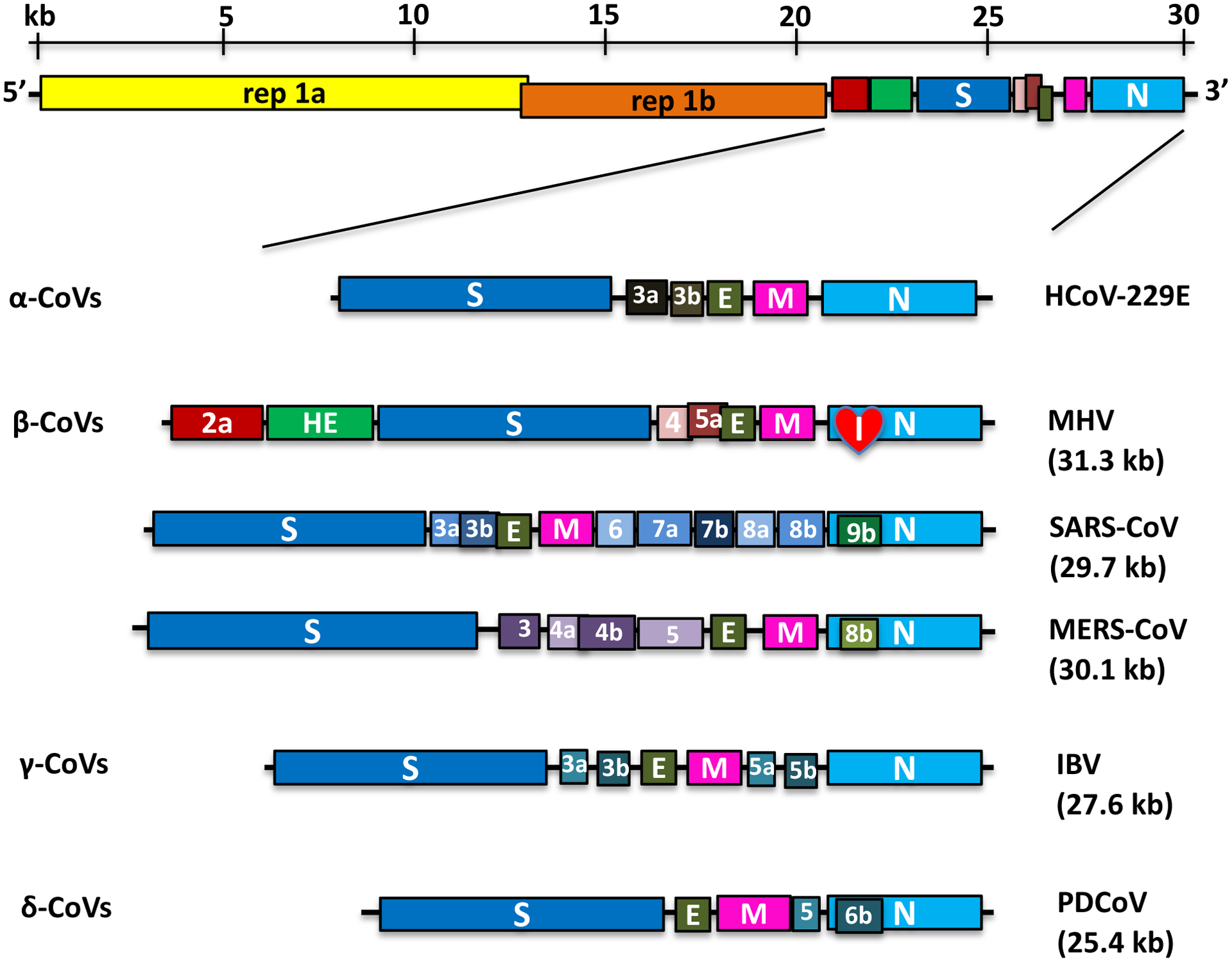
Figure 1. Genome Organization of Representative α, β, γ and δ-CoVs.

An illustration of the MHV genome is shown on top. The replicase gene constitutes two ORFs, rep 1a and rep 1b, which are expressed by a ribosomal frameshifting mechanism. The expanded regions below show the structural and accessory proteins in the 3′ regions of α-CoVs (HCoV-229E), β-CoVs (MHV, SARS-CoV, and MERS-CoV), γ-CoVs (IBV) and δ-CoVs (PDCoV). The total genome size is given for each virus. The sizes and positions of accessory genes are indicated, relative to the basic genes S, E, M, and N. The size of the genome and individual genes are approximated using the legend at the top of the diagram but are not drawn to scale.
