Fig. 1.
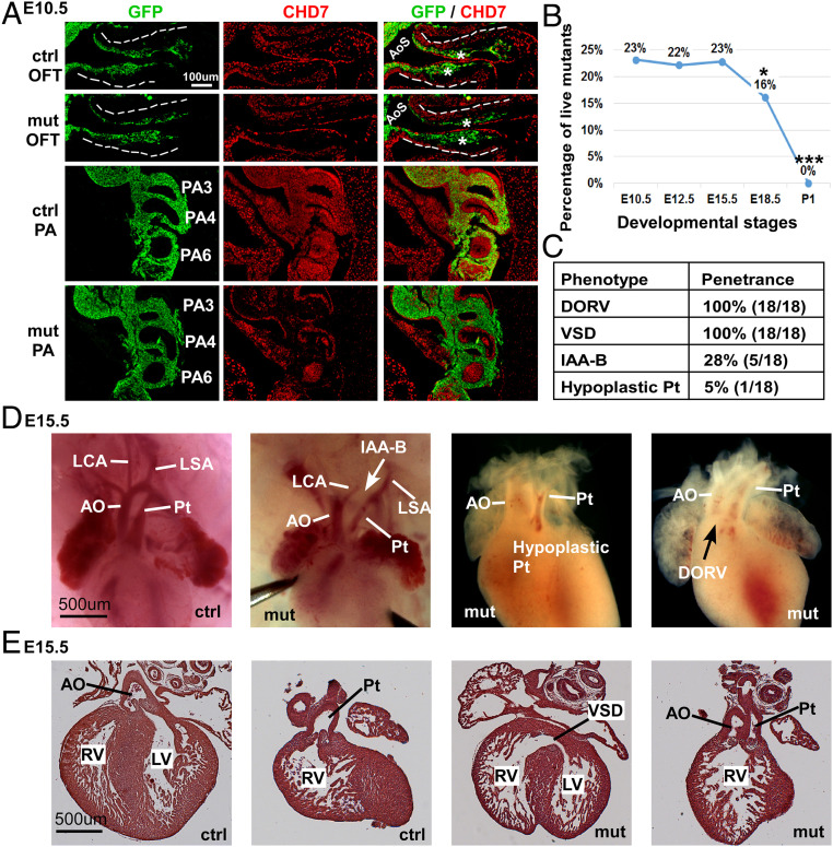
Deletion of Chd7 in NCCs led to severe conotruncal defects. (A) Immunostaining of control (Ctrl; Wnt1-Cre2;Chd7+/+;R26mtmg/+) and mutant (mut; Wnt1-Cre2;Chd7loxp/loxp;R26mtmg/+) embryos at E10.5 with antibodies against GFP (green) and CHD7 (red). Areas of the OFT and PA regions are shown. The dashed lines outline the OFT. OFT cushions are indicated with an asterisk (*). Broader views of the same embryos are shown in SI Appendix, Fig. S1C. (B) Percentage of live mutant (Wnt1-Cre2;Chd7loxp/loxp) animals, resulting from crossing Wnt1-Cre2;Chd7loxp/+ and Chd7loxp/loxp mice. Data were obtained from at least four litters at each of the indicated developmental stages. *P < 0.05; ***P < 0.001, χ2 test. (C) Summary of heart defects observed in 18 mutant embryos (Wnt1-Cre2;Chd7loxp/loxp) at E15.5. Pt, pulmonary trunk. (D) Whole-mount examination revealing conotruncal defects in mutant embryos at E15.5. AO, aorta; LCA, left carotid artery; LSA, left subclavian artery. (E) Section examination showing the DORV defect in a mutant embryo at E15.5. LV, left ventricle; RV, right ventricle.

