Fig. 4.
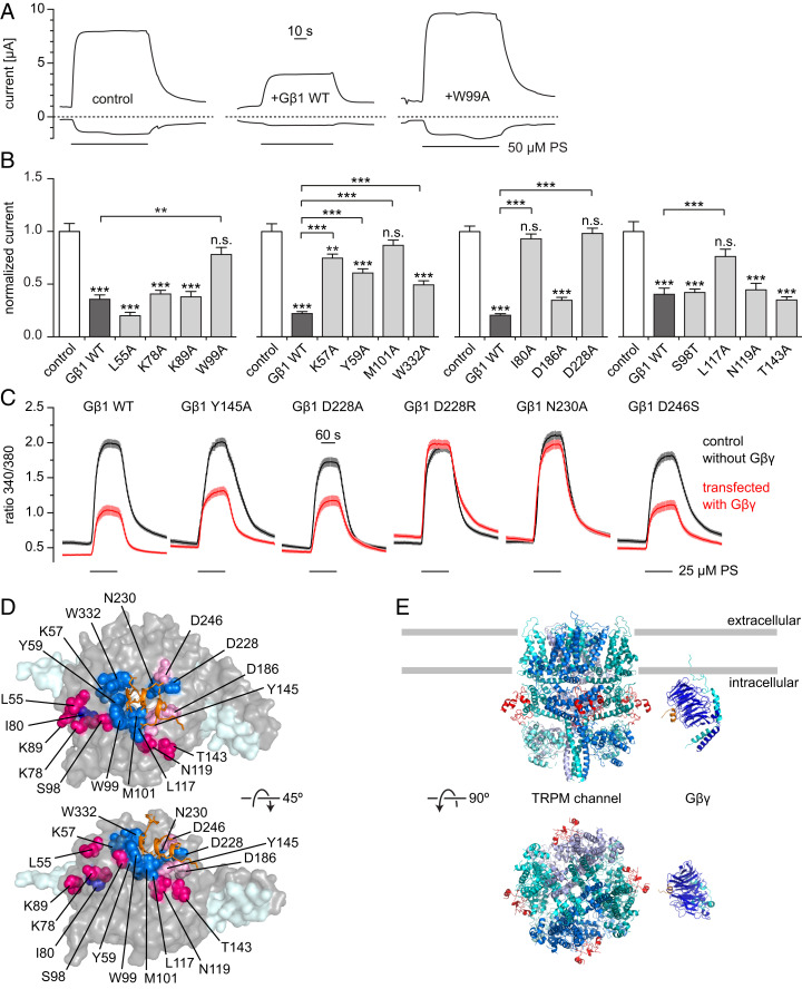
Effects of Gβ mutations on the inhibition of TRPM3. (A) Representative current traces (at −100 and +100 mV) of two-electrode voltage clamp experiments performed in Xenopus oocytes expressing hTRPM3, Gγ2 and wild-type or mutated Gβ1; 50 μM pregnenolone sulfate (PS) was applied as indicated, dashed lines denote zero current. (B) Summary data showing current amplitudes normalized to the average current induced by 50 μM PS in control oocytes without Gβγ coexpression in the same experiment. Mutants tested in the same experiments were grouped into the same panel. Asterisks above columns indicate significant difference from control oocytes without Gβγ expression (*P < 0.05; **P < 0.01; ***P < 0.001; n.s., P ≥ 0.05). (C) Ca2+ imaging experiments in TRPM3-expressing HEK293 cells transiently transfected with Gγ2-IRES-GFP and Myc-tagged Gβ1 (wild-type or mutant, red) or empty vectors as controls (black). When using the mutants D228R and N230A, the strong reduction in PS-induced Ca2+ signals seen with wild-type Gβ was not observed. SI Appendix, Fig. S7 shows single cell responses for the data in A–C, as well as expression controls for mutant Gβ1 proteins. (D) Visual summary of the Gβ mutagenesis experiments. Residues that reduced or abolished the inhibition of TRPM3 when mutated are shown as blue spheres, the TRPM3-encoded peptide is drawn as orange sticks. Residues not having an effect on TRPM3 inhibition when mutated are shown as red or pink spheres depending on the apparent distance to the TRPM3 peptide. (E) Cartoon of the proposed model: Gβγ binds to the linker region between MHR3 and MHR4 (red) located on the outer surface of the channel and at a suitable distance from the plasma membrane for binding to membrane-bound Gβγ. Since a 3D structure for TRPM3 is not available, the closely related TRPM7 [PDB ID code 6BWF (49)] is depicted (see SI Appendix, Fig. S8C for additional TRPM structures). The Gβγ structure (blue and cyan) with the TRPM3-encoded peptide (orange) is the 3D structure described in this paper. Gray bars approximately indicate the plasma membrane boundaries.

