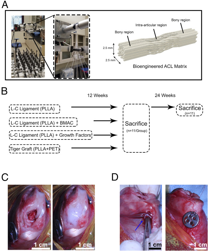
Fig. 1.
Fabrication of the bioengineered ACL matrix and implantation in a rabbit ACL reconstruction model. (A) Depiction of the braiding machine used to fabricate the bioengineered ACL matrices and the resulting biphasic structure of the matrix. Each matrix was composed of 24 yarns. (B) For the L-C ligament, 24 yarns of PLLA were braided together. For the tiger graft, 20 yarns of PLLA and 4 yarns of PET were braided together. Experimental groups were evaluated at 12 wk, and the L-C ligament (control) was further evaluated at 24 wk. (C) View of implanted bioengineered ACL matrix at the time of surgery (Left) and the application of fibrin glue (Right). BMAC or growth factors were mixed with fibrin glue for the experimental groups. (D) Representative image demonstrating the implantation of a fibrin gel in the tibial bone tunnel (Left, blue arrow) and subsequent fixation of a titanium suture button (Right).

