Fig. 3.
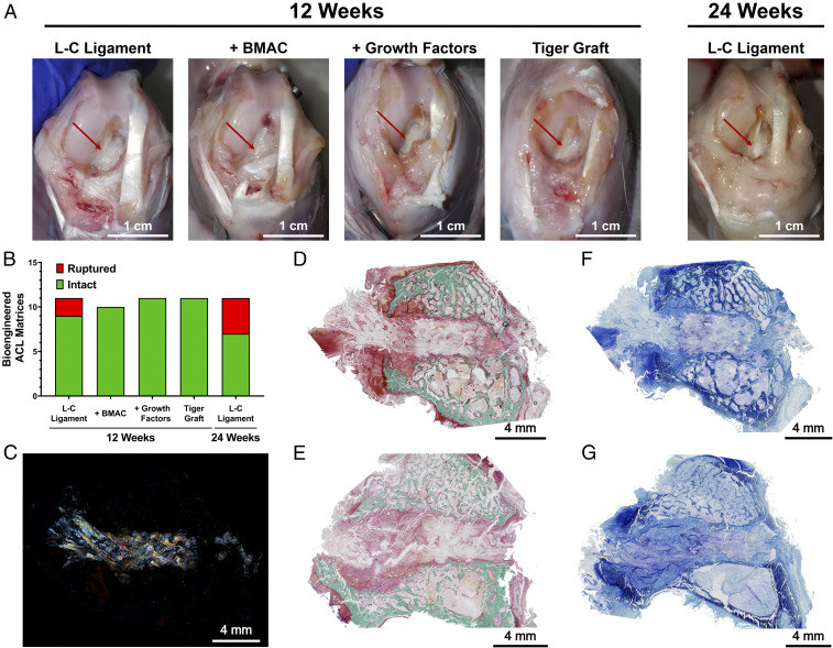
Ninety-five percent of the bioengineered ACL matrices were intact at 12 wk. (A) Representative images of knee joint and bioengineered ACL matrices (red arrow) in each group at 12 and 24 wk. (Scale bars, 1 cm.) (B) Assessment of gross state of bioengineered ligaments at 12 and 24 wk (n = 11; one BMAC sample excluded due to surgical complication); no significant difference between groups, Fisher’s exact test. (C) Polarized microscopy image demonstrates presence of braided bioengineered ligament within the bone tunnel. (D) Same histological section as C stained with Goldner’s tichrome staining. Representative Goldners Trichrome staining of (D) femoral and (E) tibial bone tunnel at 12 wk (n = 3; green, mineralized bone; scarlet, osteoid; red, soft tissue). (Scale bars, 4 mm.) Representative toluidine blue staining of (F) femoral and (G) tibial bone tunnels (n = 3; metachromatic staining, purple). (Scale bars, 4 mm.)

