Fig. 4.
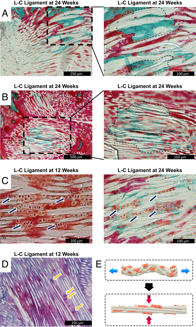
Progressive bone regeneration through endochondral ossification is seen in the L-C ligament group at 24 wk. (A) Representative image of the bioengineered ACL matrix–bone interface (n = 3, 24 wk post-ACL reconstruction). The black outlined box indicates area of higher magnification. The black outlined area represents areas with PLLA fibers embedded in mineralized bone. (B) Representative image of the L-C ligament in the bone tunnel proper (n = 3, 24 wk post-ACL reconstruction). The black outlined box indicates area of higher magnification. The black outlined area represents areas with PLLA fibers embedded in mineralized bone. (C) Representative image of the L-C ligament at 12 and 24 wk (n = 3). The navy arrows denote areas with columnar arrangement of cuboidal cells. (D) Representative Safranin O/azophloxin staining of the L-C ligament. The yellow arrows point to cuboidal cells with metachromasia staining. (E) Schematic of mechanical forces placed on bioengineered ACL matrix.

