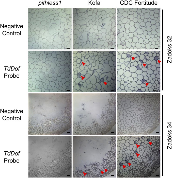
Fig. 4.
In situ PCR demonstrating enrichment of TdDof messenger RNA in internode parenchyma cells in pithless1, Kofa, and CDC Fortitude. Purple staining (labeled by red arrows) indicates the presence of in situ PCR amplified TdDof transcripts. Representative micrographs of internode cross-sections from Zadoks stages 32 and 34 show similar high expression levels of TdDof in parenchyma cells in which the negative controls were generated under the same conditions but with the reverse transcription steps omitted. Three panels from Left to Right represent internode tissues from pithless1, Kofa, and CDC Fortitude. (Scale bars = 200 μm.)

