Fig. 5.
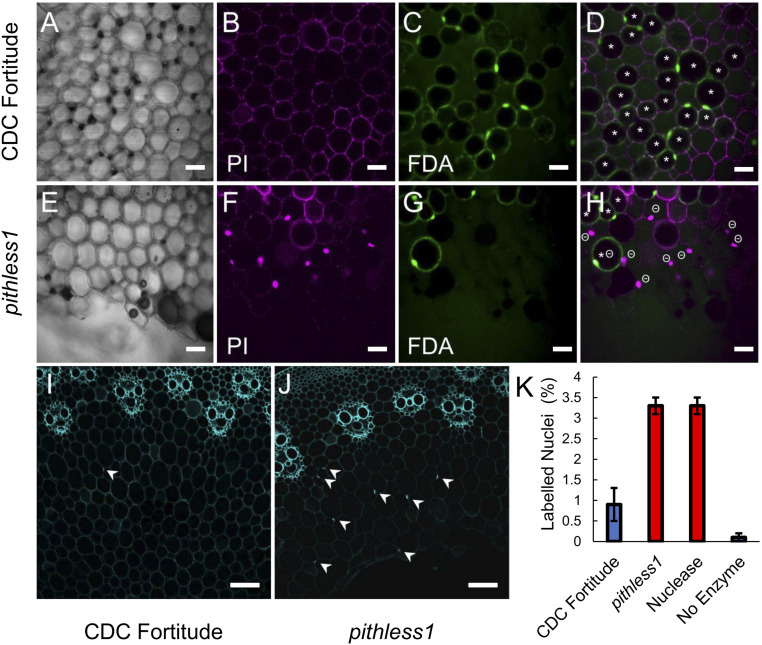
Cell viability in the pith parenchyma cells of CDC Fortitude and pithless1 stems. Stem cross-sections from the second internodes of CDC Fortitude (A–D and I) and mutant pithless1 (E–H and J) plants viewed as single frame images. Viability staining of pith cells viewed by confocal laser-scanning microscopy phase contrast image (A and E), PI signal (B and F), FDA signal (C and G), and PI and FDA merged view (D and H). Parenchyma cell walls stained with PI in CDC Fortitude (B) and pithless1 (F), with fluorescent nuclei indicating penetration of the dye into cells surrounding the hollow pith of pithless1 stem sections. Penetration and conversion of nonfluorescent FDA stain into green fluorescent fluorescein by CDC Fortitude (C) and pithless1 (G) pith parenchyma cells. The * symbol indicates a putatively viable cell, with bright FDA signal around a dark vacuole, and the Θ symbol indicates a cell considered to be dead, with PI-positive nucleus. The viability status of cells without labels is undetermined. TUNEL assay for DNA fragmentation in CDC Fortitude (I) and pithless1 (J) stems, as viewed by confocal laser scanning microscopy. Arrowheads indicate positive nuclear signals in pith cells. (K) Quantification of TUNEL assay results for three biological replicates of CDC Fortitude (Fort), pithless1 (P1), positive control using TACS nuclease treatment (+), and unlabeled experimental control (−), with the labeled nuclei per the total number of pith parenchyma cells plotted as the mean percentage ±SD. (Scale bars: A−H = 20 µm; I and J = 100 µm.)

