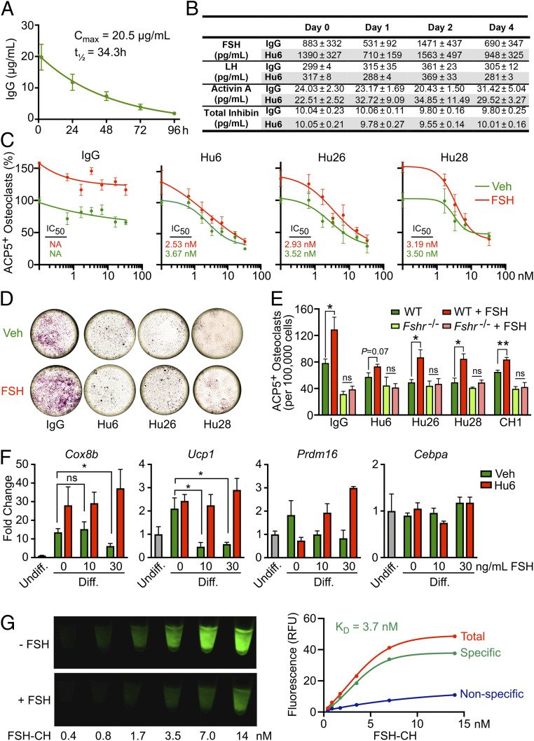
Fig. 4.
Pharmacokinetic and functional studies on humanized antibodies. (A) Plasma Cmax (20.5 µg/mL) and half-life (t1/2 = 34.3 h) of Hu6 (100 µg per mouse) injected intraperitoneally into female C57BL/6 mice (n = 6). (B) There was no difference in serum FSH, LH, activin A, or total inhibin levels in female C57BL/6 mice following one or two injections of Hu6 (100 µg per mouse; n = 5 mice). Full concentration–response curves (C) and representative light micrographs for the highest antibody concentration (66 nM; D) displaying the inhibition of ACP5-positive osteoclast formation in bone marrow cell cultures (50 ng/mL RANKL and 20 ng/mL MCSF) in response to Hu6, Hu26, or Hu28 with or without human FSH (50 ng/mL). Calculated IC50s are shown. (E) Inhibition by Hu6, Hu26, and Hu28 (6.6 nM) of FSH-induced osteoclastogenesis in wild type mice (with intact FSHRs) was not seen in cultures from Fshr−/− mice, proving that the antibodies act via the FSH axis. (F) Expression of beiging genes, namely Cox8b, Ucp1, Prdm16, and Cebpa, in response to Hu6 (6.6 nM) and/or FSH (10 or 30 ng/mL) in 3T3.L1 cell cultures under differentiating conditions (Methods). (G) 3T3-L1 cells grown in differentiation medium with 10 µM troglitazone for 8 d were incubated with FSH-CH for 2 h (37 °C) with or without a 100-fold molar excess (1.4 µM) of nonconjugated FSH. Cells were scanned using Li-Cor Scanner (800-nm channel). Scanned images (Left) and saturation curves based on quantitation of scanned images (Right) are shown (KD = 3.7 nM).

