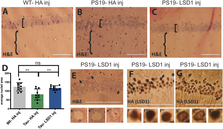
Fig. 5.
LSD1 overexpression rescues the neurodegenerative phenotype in the hippocampus of 11-mo-old PS19 Tau mice. (A–C) Representative image of H&E-stained CA1 region of the hippocampus of 11-mo-old wild-type mice injected with HA control virus (WT-HA inj) (A), PS19 Tau mice injected with HA control virus (PS19-HA inj) (B), and PS19 Tau mice injected with Lsd1 overexpressing virus (PS19-LSD1 inj) (C). Square brackets denote thickness of pyramidal layer of the CA1 of the hippocampus and braces denote hippocampal region with or without infiltrating cells. (D) Quantification of the average number of nuclei in the pyramidal layer of the hippocampus per area per mouse from histology represented in A–C. Values are mean ± SD (n = 10, one-way ANOVA with Tukey’s post hoc test, **P < 0.01; ns, not significant). (E) Representative H&E of PS19-LSD1 inj mouse with abnormal nuclei blebbing in the CA1 region of the hippocampus. E′–E‴, high magnified image of cells denoted by arrows in E of individual nuclei that are either abnormally blebbed (E′, E″) or normal (E‴). (F and G) Immunohistochemistry staining of HA(LSD1) in 11 mo PS19-LSD1 inj mice. HA is either localized specifically to the nucleus in all nuclei (F) or in only a few nuclei while it is partially sequestered in the cytoplasm in others (G). F′–F‴, high magnified image of cells denoted by arrows in F of nuclear HA localization in individual nuclei. G′ and G‴, high magnified image of cells denoted by arrows in G of individual nuclei with HA(LSD1) either sequestered to the cytoplasm (G′, G″) or confined to the nucleus (G‴). (Scale bars, 50 μm.)

