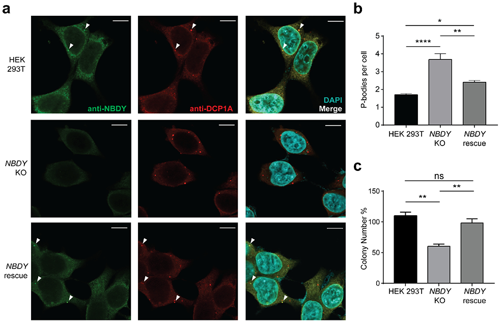
Figure 2∣. Endogenous NBDY localizes to and regulates P-bodies and affects cell proliferation.

(a) Detection of endogenous NBDY by immunofluorescence. Fixed cells were stained with antibodies detecting NBDY and a P-body marker, DCP1A. (b) P-body numbers in HEK 293T, NBDY KO and NBDY rescue cell lines. Six fields of view for each cell line (>180 cells) were used to quantify average P-bodies (anti-DCP1A, P-body marker) per cell. Data represent mean ± s.e.m, and significance was evaluated with one-way ANOVA. *P < 0.05; **P < 0.01; ****P < 0.0001, Dunnett’s test. Scale bars, 10 μm. For additional field of view, see Figure S1a. (c) Cell proliferation assay for HEK 293T, NBDY KO and NBDY rescue cell lines. Significance was analyzed by one-way ANOVA. **P < 0.01, Dunnett’s test.
