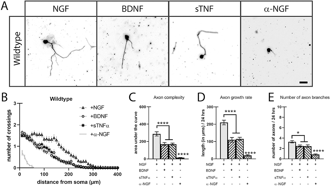
Fig. 1.
Soluble BDNF and TNFα reduces axon complexity in cultured sympathetic neurons.
A) SCG neurons were cultured in the presence of NGF (2 ng/mL) or a combination of NGF with p75NTR ligand BDNF (250 ng/mL), TNFR1 ligand sTNFα (2 ng/mL), or NGF-neutralizing antibody α-NGF (2 ng/mL) and stained with TUJ1 24 h after plating. Scale bar = 30 μm. B) Sholl analysis of P0 SCG neuron axon complexity was performed by counting trace intersections in 10 μm intervals. C) Quantification of the area under the curve from B). D) Axon growth rate was measured as the length of the longest axon segment per neuron after 24 h from B). E) Axon branching was measured as the number of axon branch endpoints per neuron after 24 h from B). Full statistical comparisons found in STable 1. Error bars represent s.e.m. n = 62 to 112 neurons per condition from 3 cultures of SCGs pooled from whole litters. *p < 0.05, ****p < 0.0001 using 2-way ANOVA with Tukey’s multiple comparisons test.

