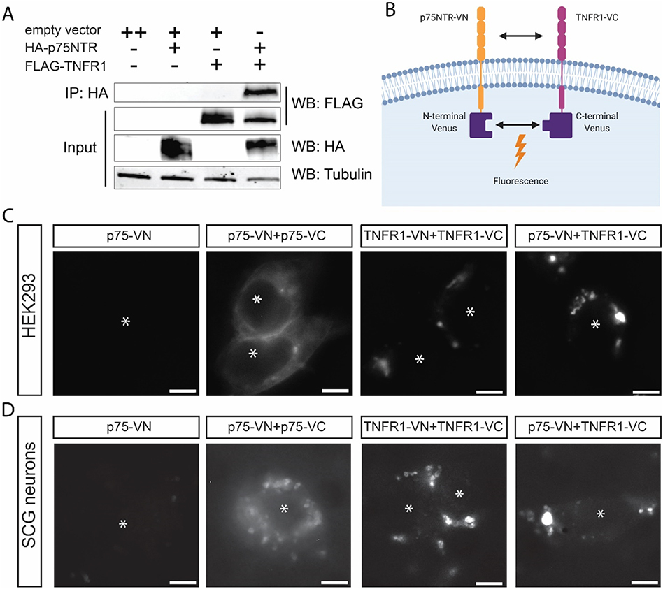
Fig. 3.
TNFR1 interacts with p75NTR and changes its subcellular localization.
A) Co-immunoprecipitation (co-IP) of p75NTR and TNFR1 constructs using HEK293 cells. The experiment was performed 4 times (n = 4). B) Schematic of bimolecular fluorescence complementation (BiFC). This figure was created with Biorender.com and exported under a paid subscription. C, D) Bimolecular fluorescence complementation (BiFC) of p75-NTR and TNFR1 constructs using HEK293 cells (C) or sympathetic neurons (D). Transfected cells were imaged using fluorescence excitation at 514 nm. Asterisks indicate the cells in each image. Images are representative of experiments performed at least 3 times (n = 3). Scale bar = 5 μm.

