
Figure 6.
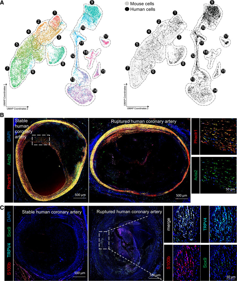
Human scRNA-seq shows similar groups of fibrous cap and osteogenic SMC in advanced lesions. A, UMAP analysis of scRNA-seq results from human carotid endarterectomy samples, colored by cluster (left) or by origin (right) against mouse scRNA-seq data from Figure 2. B, Human coronary artery lesions graded as stable or ruptured were immunostained for Acta2 and Phactr1, a marker of cluster 1 to 3 SMCs identified in our scRNA-seq analyses of advanced BCA lesions. Images of whole immunostained human coronary lesions were obtained by taking a tile scan of ×20 zoom confocal microscopy z-stacks of full 10-µm thickness. C, Human coronary artery lesions graded as stable or ruptured were immunostained for Sox9, TRPV4, and S100b, which were markers of osteogenic SMCs identified in our scRNA-seq analyses of mouse advanced BCA lesions, imaged as described in B. BCA indicates brachiocephalic artery; scRNA-seq, single-cell RNA sequencing; SMC, smooth muscle cell; and UMAP, uniform manifold approximation and projection.