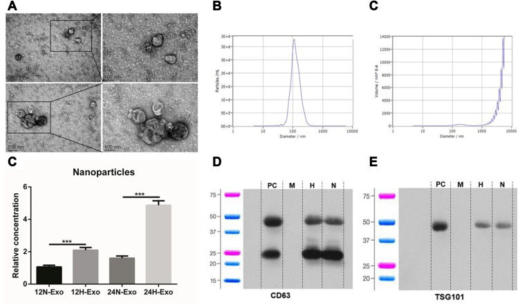
Figure 1.
Isolation and identification of exosomes. (A) Exosomes isolated from the supernatants of Eca-109 cells under TEM. (B and C) NTA of exosomes particle concentration and particle size distribution. (D) Relative concentration of nanoparticles in 12N, 12H, 24N and 24H (***p<0.01). (E and F) Western blot analysis for exosomal proteins CD63 and TSG101.
Abbreviations: PC, positive control; M, marker; H, hypoxia; N, normoxia.

