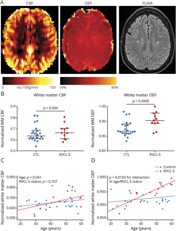
Figure 4. White matter OEF increases with disease duration and may indicate progressive microvascular ischemia.
(A) Representative cerebral blood flow (CBF), oxygen extraction fraction (OEF), and fluid-attenuated inversion recovery (FLAIR) maps in an individual with retinal vasculopathy with cerebral leukoencephalopathy and systemic manifestations (RVCL-S). (B) Normalized white matter OEF (nOEF_NAWM), but not normalized white matter CBF (nCBF_NAWM), was significantly elevated in patients with RVCL-S compared to healthy controls (CTLs). (C) nOEF_NAWM progressively increased in participants with RVCL-S compared to CTLs (p = 0.0103); however, rate of change in nCBF_NAWM was not different in those with RVCL-S compared to CTLs.

