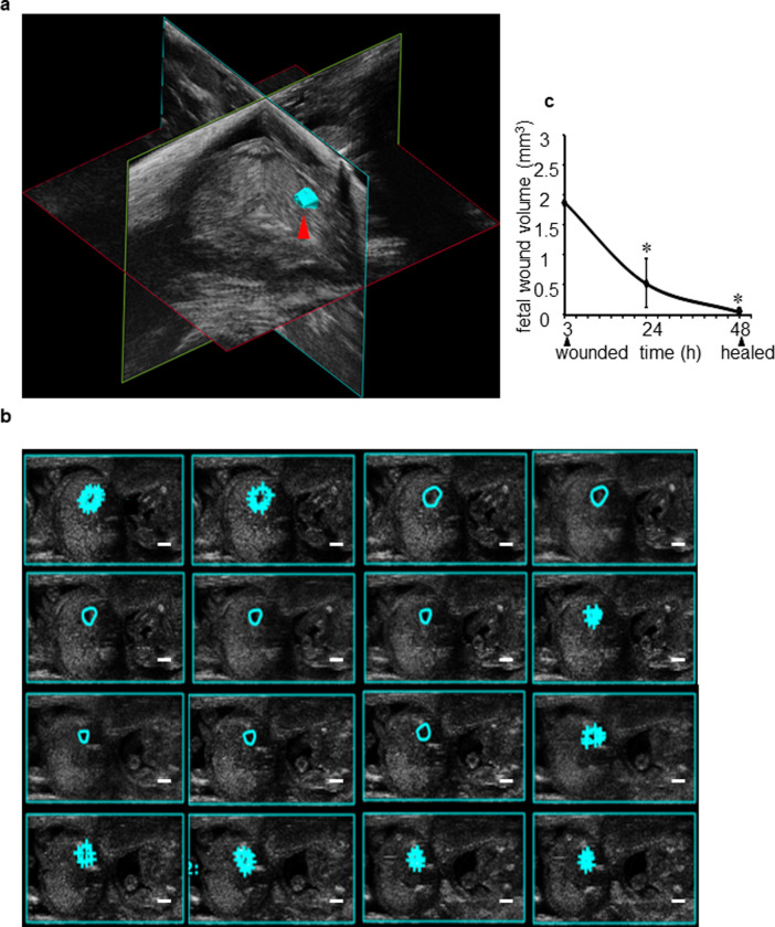
Fig 2. Volumetric fetal wound measurement using three-dimensional (3D) reconstruction of ultrasound images.
(a) A 3D view of fetal wound (cyan) surface rendered 3D object constructed from 16 contours structure of fetal wound using the ultrasound B-mode images as shown by red arrow. (b) Semi-automated tracing of fetal wound border through different frames obtained from 3D visualization to measure wound volume (cyan). The tracing was performed manually in different frames of the 3D recorded video. The algorithm in ‘VevoStrain’ package then automatically traced the wound boarders on the frames that are in between the manually traced frames. (c) Line graph plot showing change in fetal wound volume over time at 3, 24, and 48h post-wounding. Data represented as the mean ± SE, n = 3 wounds. *p< 0.05.

