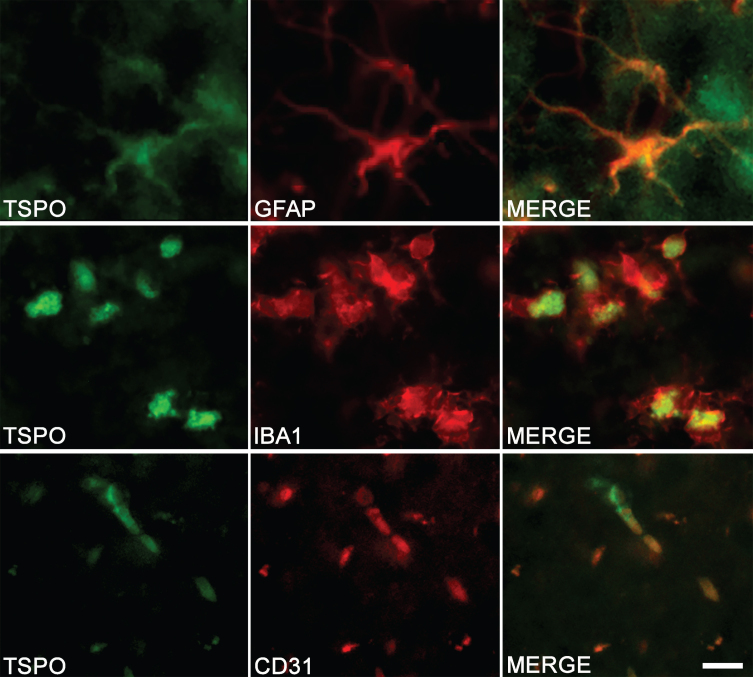
Fig. 3.
Cell origin confirmation of TSPO expression in the hippocampus of TgF344-AD rats. Double-immunofluorescence was performed with antibodies directed against TSPO (green color) and specific cell markers (red color). Merge images demonstrate the colocalization of TSPO with astrocytes (GFAP), microglia (IBA1), and endothelial cells (CD31). Scale bar: 10μm.

