Figure 1.
Confirmation of antibody specificity for PI5P4Kα and PI5P4Kβ in mouse brain
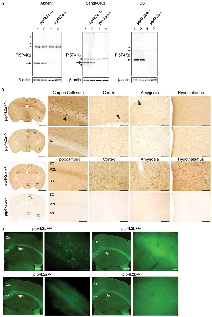
a. Western blot analysis of whole brain lysates in wild-type and pip4k2a knockout mouse brain shows specificity of 2 antibodies (Abgent and Santa-Cruz) for PI5P4Kα, and specificity of Cell Signal antibody (CST9594S) for PI5P4Kβ in wild-type compared to knockout brains. Arrows, specific bands that are not present in knockout brain lysates; *, non-specific bands.
b. Immunoperoxidase staining for PI5P4Kα (top two rows) (Abgent) and PI5P4Kβ (bottom two rows) in wild-type and knockout brains shows positive signal in wild-type brains and lack of staining in pip4k2a and pip4k2b knockout brains. Corpus callosum, cortex, basolateral amygdala, and hypothalamus are shown for PI5P4Kα staining, and hippocampus, cortex, basolateral amygdala, and hypothalamus are shown for PI5P4Kβ staining. Scale bars for immunoperoxidase images are 2 mm for low-magnification images on left and 125 μm for high-magnification images on right.
c. Immunofluorescence for PI5P4Kα (left) (Abgent) and PI5P4Kβ (right) in wild-type and knockout brains shows positive PI5P4Kα signal in corpus callosum and positive PI5P4Kβ signal in hippocampus in wild-type brains and lack of staining in pip4k2a and pip4k2b knockout brains. Scale bar length is indicated for each bar.
CC, corpus callosum; PCL, pyramidal cell layer; SO, stratum oriens; SR, stratum radiatum; Hpc, hippocampus; Ctx, cortex

