Figure 10.
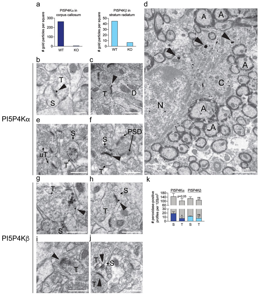
Immuno-electron microscopy of endogenous PI5P4Kα and PI5P4Kβ expression in mouse hippocampus and corpus callosum.
a Quantification of SIG particle labeling for PI5P4Kα (Abgent) in corpus callosum and PI5P4Kβ in stratum radiatum (SR) of hippocampal CA1 from wild-type and respective knockout mice.
b-d SIG particle labeling for PI5P4Kα (arrowheads) is contained in an axon terminal, adjacent to the synapse (b), the perisynaptic zone of an asymmetric synapse in a dendritic spine in CA1 SR (c), and oligodendrocyte cytoplasm in corpus callosum (d).
e-f Immunoperoxidase labeling for PI5P4Kα (arrowheads) is contained in an axon terminal (e) and dendritic spine (f) in CA1 SR. Unlabeled terminals (uT) and post-synaptic densities (PSD) are indicated for comparison.
g-h SIG labeling for PI5P4Kβ (arrowheads) is found in an axon terminal (g) and dendritic spine (h) in CA1 SR.
i-j Immunoperoxidase labeling for PI5P4Kβ (arrowheads) is found in an axon terminal (i) and dendritic spine (j).
k Quantification of PI5P4Kα and PI5P4Kβ immunoperoxidase labeled profiles in CA1 (n=3 mice per group). Labeled profiles are indicated by dark blue (PI5P4Kα) and light blue (PI5P4Kβ) bars, and unlabeled profiles are indicated by gray bars. *, p < 0.05; ns, not significant.
Scale bars for b, c, e, f, g, h, i, j = 500 nm; scale bars for D = 2 μm.
A, axon; C, cytoplasm; S, dendritic spine; N, nucleus; T, axon terminal; uT, unlabeled axon terminal

