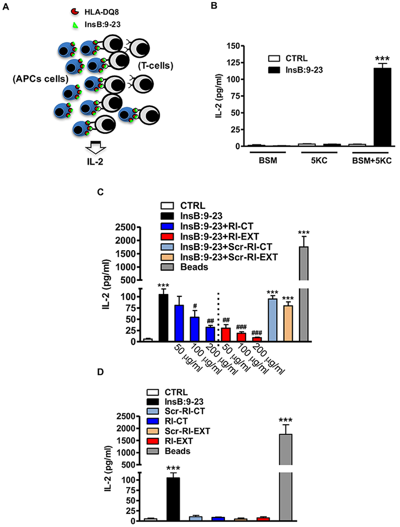
Fig. 3. RI-CT and RI-EXT inhibit the production of IL-2 in a cellular antigen presentation assay with BSM cells and 5KC cells.

(A) BSM cell line (APCs) loaded with InsB:9-23 peptide and a murine T-cell clone (T-cells) expressing a human TCR specific for the InsB:9-23–DQ8 complex (5KC cells) were used to test functionally in vitro RI-CT and RI-EXT. (B) In our system, IL-2 was detected only when BSM cells and 5KC cells were incubated together with InsB:9-23. (C) Both RI-CT and RI-EXT inhibited significantly IL-2 production compared to untreated cells, starting at a concentration of 100 μg/ml and 50 μg/ml respectively. (D) RI-D-peptides alone or their scrambled versions did not induce IL-2 production. CD3/CD28 mouse beads were used as positive control; supernatants were analyzed by Luminex for IL-2. Bars represent mean ± SEM from 4 to 5 independent experiments. ***p < 0.001, by Student’s t-test for comparison of treated cells versus control cells. ###p < 0.001; ##p < 0.01; #p < 0.001 by Student’s t-test for comparison of RI-D-peptides treated cells versus InsB:9-23 treated cells.
