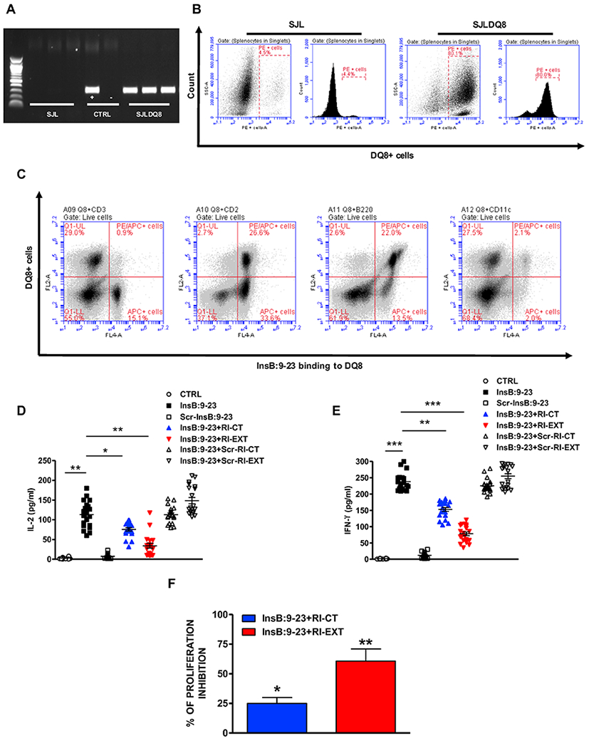
Fig. 4. Ex vivo effect of RI-CT and RI-EXT.

(A) and (B) PCR and flow analysis confirming the presence of the transgene in SJL-DQ8 mice compared to WT SJL mice. Cells were gated on live splenocytes in singlets. (C) Flow assay showing the expression of HLA-DQ8 molecule in lymphocytes isolated from SJL-DQ8 mice. HLA-DQ8 molecule is expressed mostly on APCs. Cells were gated on live splenocytes in singlets. (D-F) 20 SJL-DQ8 mice were immunized subcutaneously with InsB:9-23 in CFA on day 1 and in IFA on day 8. 9 days after the second immunization (day 17) mice were sacrificed. (D) and (E) Lymphocytes isolated from SJL-DQ8 mice were stimulated with InsB:9-23 or with scrambled InsB:9-23 as negative peptide and incubated with RI-CT or RI-EXT (scrambled RI-CT or RI-EXT were used as negative peptides). Supernatants were analyzed by Luminex for IL-2 and IFN-γ. (F) Inhibition of T-cell proliferation by RI-CT or RI-EXT was analyzed by the CFSE assay after stimulation of lymphocytes with InsB:9-23 with or without addition of RI-CT or RI-EXT. Cells were gated on live splenocytes in singlets. Both RI-D-peptides significantly decreased secretion of cytokines and proliferation induced by InsB:9-23. *p < 0.001; **p < 0.01; ***p < 0.001, by Student’s t-test.
