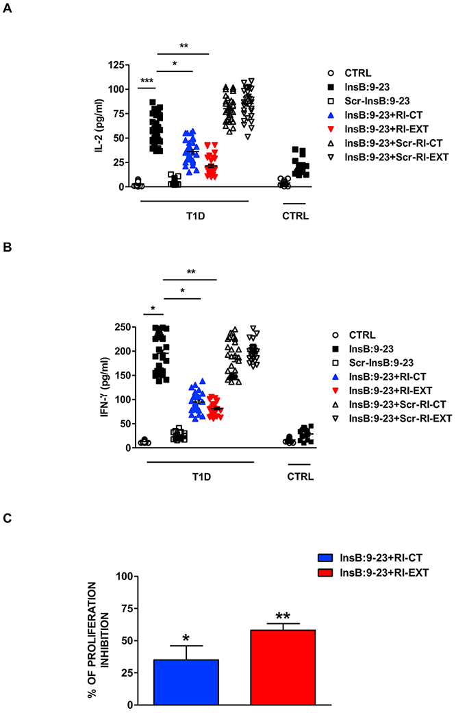
Fig. 6. Effect of RI-CT and RI-EXT in PBMCs from new onset T1D patients.

(A) and (B) PBMCs were isolated from 31 new onset DQ8 positive T1D patients and 17 DQ8 positive controls and were stimulated for 48h with InsB:9-23, with or without RI-CT and RI-EXT (scrambled InsB:9-23, scrambled RI-CT and scrambled RI-EXT were used as negative peptides). The production of IL-2 and IFN-γ were assessed by Luminex. RI-CT and RI-EXT significantly decreased T-cell activation induced by InsB:9-23, whereas scrambled RI-CT or RI-EXT had no effect. (C) Inhibition of T-cell proliferation by RI-CT or RI-EXT was analyzed by the CFSE assay after stimulation of human PBMCs with InsB:9-23 with or without addition of RI-CT or RI-EXT. PBMCs were gated on live T-cells for flow cytometry analysis. Both RI-D-peptides significantly decreased proliferation induced by InsB:9-23 in PBMCs isolated from DQ8-T1D patients. *p < 0.001; **p < 0.01; ***p < 0.001, by Student’s t-test.
