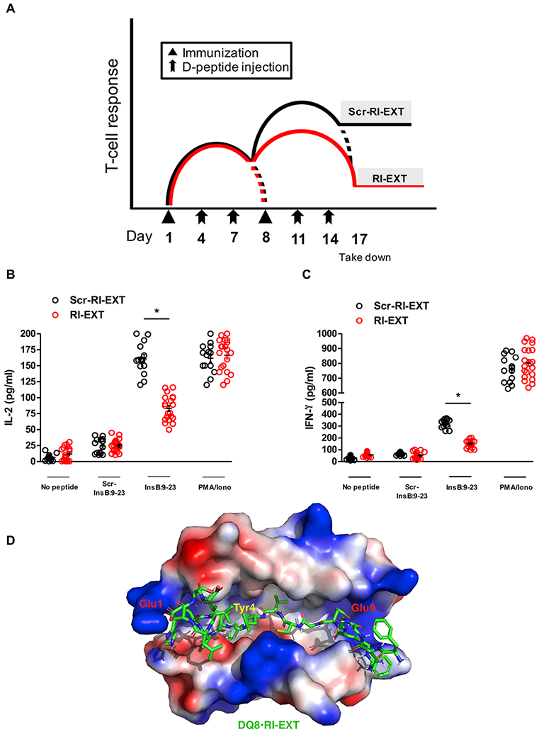
Fig. 7. RI-EXT blocks activation of T-cells to InsB:9-23 in vivo.

(A-C) SJL-DQ8 mice were immunized subcutaneously with InsB:9-23 in CFA on day 1 and with InsB:9-23 in IFA on day 8. At days 4, 7, 11 and 14 mice were injected with RI-EXT (20 mice) or scrambled RI-EXT as negative control peptide (14 mice). 9 days after the second immunization (day 17) mice were sacrificed. Lymphocytes isolated from SJL-DQ8 mice were stimulated with InsB:9-23 or with scrambled InsB:9-23 as negative peptide. PMA/Ionomycin was used as positive control. (B) and (C) Supernatants were analyzed by Luminex for IL-2 and IFN-γ. RI-EXT significantly blocked the InsB:9-23 induced activation of T-cells in InsB:9-23-immunized mice. There was no significant decrease of T-cell activation in response to scrambled RI-EXT. *p < 0.001, by Student’s t-test for comparison of RI-EXT versus scrambled RI-EXT treated mice. (D) Molecular model of the HLA-DQ8 binding cleft and RI-EXT; the critical residues Glu1, Tyr4 and Glu9 are highlighted.
