Fig. 7.
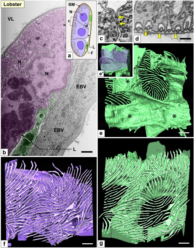
Nephrocytes in lobster (I): FIB-SEM sectional and reconstruction images. (a) Schematic drawing. Multiple nephrocytes (N) form an epithelial sac with a narrow lumen (L). The sac is partially altered into flat cells (F, green cells). There is no cytoplasmic continuity between nephrocytes. (b–d) FIB-SEM sectional images. (b) Connecting part of the nephrocytes (purple) and flat cells (green), which are in contact with the internal wall of the efferent branchial vessel (EBV). (c, d) The cell bodies and primary processes of adjacent nephrocytes were closely apposed and connected via spotty intercellular junctions (arrows in c), which were structurally different from the slit diaphragm between foot processes (arrowheads in d). (e–g, e′) Reconstruction images of two adjacent nephrocytes (green and purple). (e) Luminal view of the green nephrocyte showing its cell body was divided into three massive parts (asterisks). (e′) The green and purple cells interdigitated each other by these massive parts. (f, g) Basal view of purple and green nephrocytes. Numerous fine, long foot processes (whitish-purple in f, whitish-green in g) protruded from each massive part. Scale bars, 2 μm in b; 100 nm in c–g. The reconstructed nephrocytes (e–g) are also shown in Supplementary Movie S7

