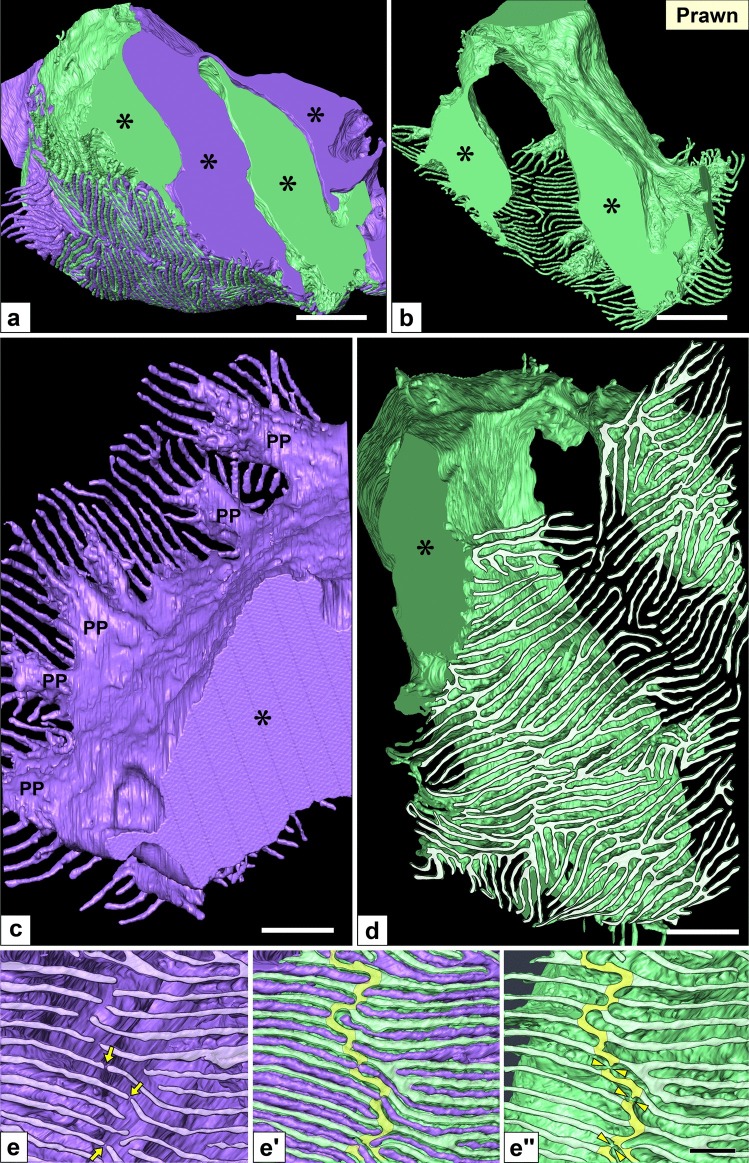
Fig. 9.
Nephrocytes in prawn (II): reconstruction images. (a–d) Reconstruction images of two adjacent nephrocytes (green and purple). (a, b) The cell body was divided into two massive parts (asterisks), while typically it is horseshoe-shaped (b). The massive parts were interdigitated between adjacent purple and green nephrocytes (a). (c) Luminal view of a massive part of the purple nephrocyte. Numerous fine long foot processes protruded from the massive part, some of which protruded via short thick primary processes (PP). (d) Basal view of the green nephrocyte, numerous fine long foot processes (whitish-green) protruded from each massive part. (e–e″) Magnification of the basal surface. Foot processes protruded via ridge-like prominences (RLP) (yellowish-green). RLP was partially retracted (arrowheads in e″). Thus, foot processes of the purple nephrocyte were closely apposed across the retracted RLP (arrows in e). Scale bars, 1 μm in a, b; 500 nm in c, d; 100 nm in e–e″. The reconstructed nephrocytes (a–d) are also shown in Supplementary Movies S8 and S11

