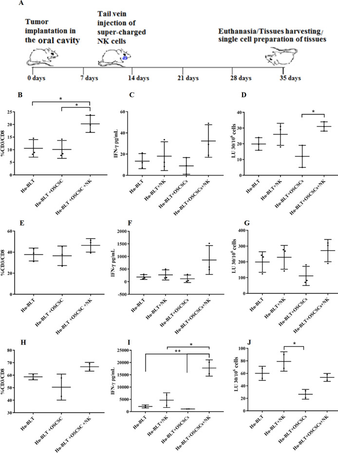
Figure 6.
OC-expanded NK cell immunotherapy increased CD8 + T cells, IFN-γ secretion, and NK cell-mediated cytotoxicity in BM, spleen, and peripheral blood of hu-BLT mice. Hu-BLT mice were orthotopically injected with 1 × 10⁶ human OSCSCs into the floor of the mouth. One to two weeks after the tumor implantation, mice received OC-expanded NK cells via tail-vein injection. The disease progression and weight loss were monitored for another 3–4 weeks (n = 3) (A). Hu-BLT mice were implanted with OSCSC tumors and were injected with NK cells as depicted in Fig. 6A. At the end of experiment, hu-BLT mice were sacrificed; the spleens, BM, and peripheral blood were harvested; and single cell suspensions were obtained and cultured as described in the Materials and Methods section. Surface expressions of CD3 and CD8 were analyzed on day 7 of BM cultures (n = 3) (B), spleen cultures (n = 3) (E), and PBMC cultures (n = 2) (H) using flow cytometry. The supernatants were harvested from the cultures on day 7 of BM culture (n = 3) (C), spleen culture (n = 3) (F), and PBMC culture (n = 2) (I), and the secretions of IFN-γ were determined using single ELISA. Cytotoxicity of day 7 cultured BMs (n = 3) (D), spleens (n = 3) (G), and PBMCs (n = 2) (J) were determined against OSCSCs using standard 4-h 51Cr release assay. LU 30/106 cells were determined using the method described in Fig. 1B.

