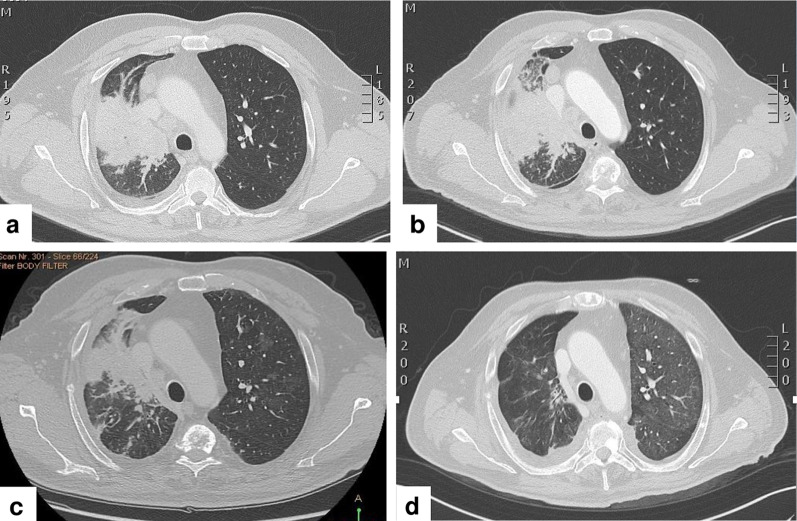
Fig. 1.
a Computed tomography scan showing solid tissue localized at the lung hilum that infiltrates the right upper lobe bronchus with concomitant atelectasis of pulmonary parenchyma. b Progression of the solid tissue with an increase in atelectasis after one cycle of pembrolizumab. c Reduction in both the size of solid tissue and pulmonary parenchyma after two cycles of chemotherapy. d Further reduction in the size of solid tissue with absence of atelectasis after 3 weeks of treatment with the RET (rearranged during transfection) inhibitor pralsetinib

