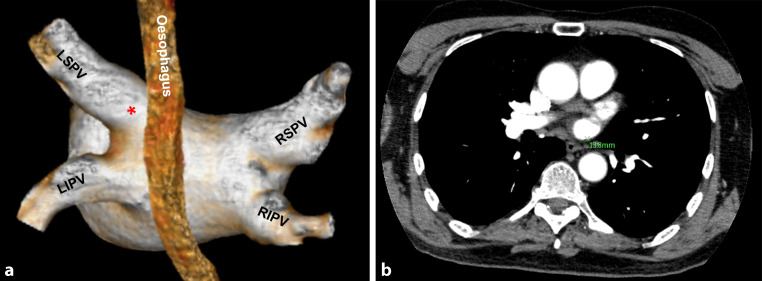
Fig. 2.
a Shortest oesophagus to pulmonary vein distance measurement when we first looked at the CT images. The origin of the os, identified as the indentation in the posterior wall, caused by the angulation of the pulmonary vein compared to the atrial wall. The angulation between the pulmonary vein and the atrium can be well distinguished in this 3D view. The distance from the identified angulation (*) to the oesophagus in the axial plane represents the shortest oesophagus to pulmonary vein distance. LSPV left superior pulmonary vein, RSPV right superior pulmonary vein, LIPV left inferior pulmonary vein, RIPV right inferior pulmonary vein. b This distance was measured

