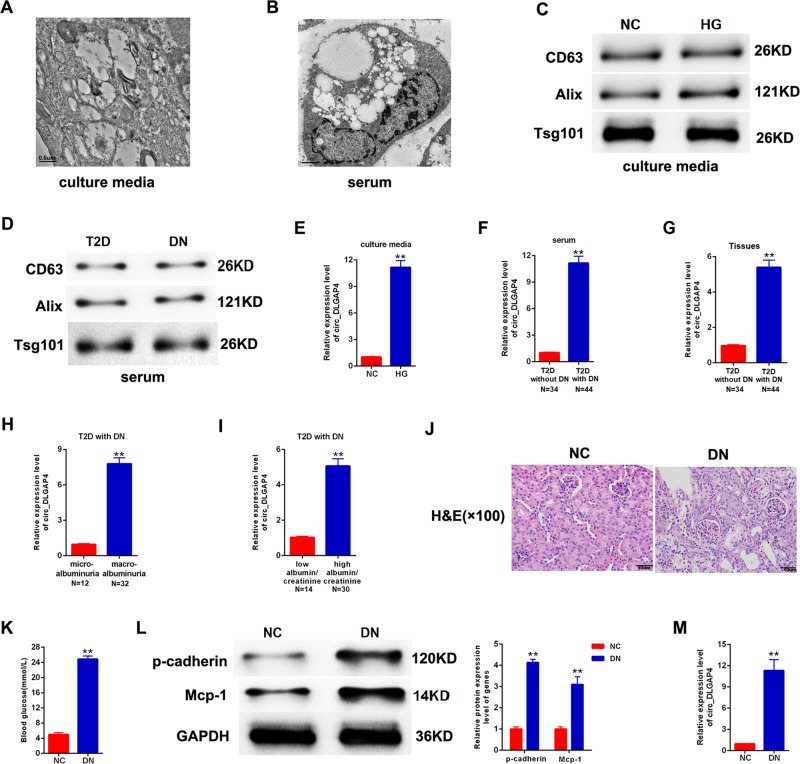
Fig. 1. The expression patterns of exo-circ_DLGAP4 in DKD.
A Scanning of exosomes isolated from culture media of MCs using electron microscopy. B Scanning of exosomes isolated from human serum using electron microscopy. C CD63, Alix, and Tsg101 protein expression in culture media of MCs was detected using western blotting analysis. D CD63, Alix, and Tsg101 protein expression in human serum was evaluated using western blotting analysis. E The expression level of circ_DLGAP4 was detected in exosomes isolated from culture media of MCs using qRT-PCR. MCs were treated with NG (5.5 mM) or HG (25 mM) for 24 h. F The expression level of circ_DLGAP4 was tested in exosomes isolated from human serum samples of T2D and DKD patients. G The expression level of circ_DLGAP4 was detected in human serum samples of T2D patients without DKD (n = 34) or with DKD (n = 44). H Expression levels of circ_DLGAP4 was detected in DKD with microalbuminuria (n = 12) or with macroalbuminuria (n = 32). I The expression level of circ_DLGAP4 were detected in DKD with low eGFR (n = 14) or high eGFR (n = 30). J HE staining in NC and DKD rat model. n = 8 in each group. K Blood glucose was detected in NC and DKD rat model. L Western blotting analysis of p-cadherin and Mcp-1 in NC and DKD rat model. M The expression level of circ_DLGAP4 was detected in NC and DKD rat model. Three independent experiments were conducted. Error bars stand for the mean ± SD of at least triplicate experiments; **P < 0.01.

