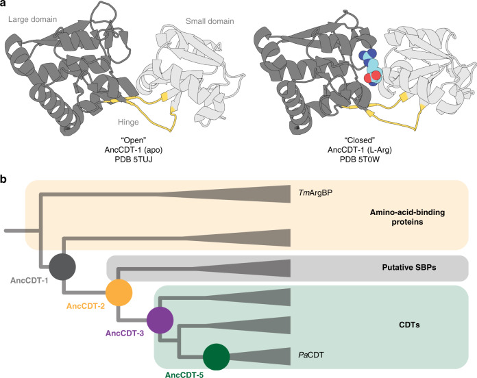
Fig. 1. The conformational change of SBPs and the evolution of PaCDT.
a X-ray crystal structures of SBPs that are specialised for binding solutes, such as AncCDT-1 (shown), typically capture open ligand-free (left, PDB 5TUJ) and closed liganded (right, PDB 5T0W) states. b Schematic drawing (not to scale) of the phylogenetic tree used for ancestral sequence reconstruction in Clifton et al.40, which highlights the evolutionary relationship between the polar amino acid-binding proteins (e.g., Thermotoga maritima L-arginine-binding protein, TmArgBP), AncCDT-1, AncCDT-3, AncCDT-5 and PaCDT. Clades are collapsed. Figure adapted from Clifton et al.40.

