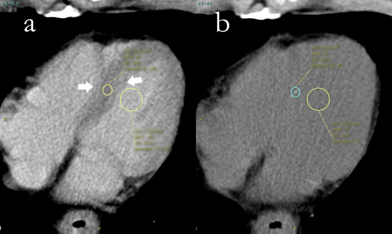
Fig. 1.
Region of interest placement for extracellular volume calculation in computed tomography of a 65-year-old male patient with adenocarcinoma of lower esophagus. a Contrast-enhanced scan, where papillary muscles are visible and thus avoided. The septum is clearly distinguishable from the intraventricular blood pool, and a certain degree of blurring is noticeable toward the borders (white arrows). b Unenhanced scan. Myocardial tissue is almost unrecognizable from the intraventricular blood pool, and regions of interest are placed in roughly the same position of a and then adjusted following local attenuation measurement

