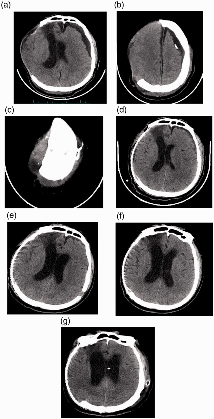
Figure 1.
Representative computed tomography (CT) scans of a 47-year-old male patient that was admitted to hospital 1 month after a previous head trauma with decompressive craniectomy and deterioration of conscious state following an apparent good recovery. (a) CT scan after the patient was admitted to hospital showing a contralateral subdural effusion (SDE) and midline shift to the side of the cranial defect, with brain tissue herniation. (b & c) CT scans after the patient was admitted to hospital showing the Ommaya reservoir that had been implanted during week 4 after the initial head injury. (d) CT scan after the cranioplasty showing the reduced contralateral SDE. (e) CT scan 9 days after the cranioplasty showing that the contralateral SDE had totally disappeared. In this image, there are also signs of hydrocephalus with mild interstitial oedema appearing around the anterior horn of the left lateral ventricle; but the patient appeared to be well without any symptoms of hydrocephalus. (f) CT scan 2 weeks after the cranioplasty showing apparent hydrocephalus. The patient had the symptoms of an uncontrolled bladder. (g) CT scan after the successful ventriculoperitoneal shunt procedure.

