Figure 1.

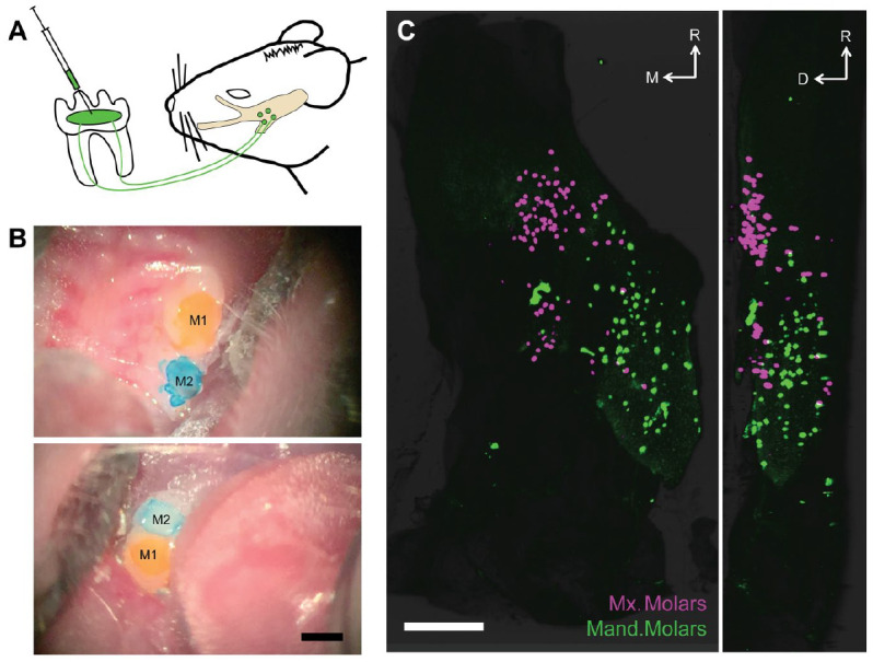
Retrograde labeling of the trigeminal sensory neurons reveals rich and selective innervation of molar teeth. (A) Schematic depicting our labeling paradigm: briefly, we prepared shallow surgical cavitations in the mouse molars, applied retrograde tracer, and observed robust labeling in somata of trigeminal sensory neurons from 16 to 72 h after labeling. (B) Images of maxillary (top) and mandibular molars (bottom) after application of distinct retrograde tracers CTB-488 (yellow) and CTB-647 (blue) and placement of dental composite. Scale bar: 500 μm. CTB, cholera toxin B-subunit. M1 and M2 denote first and second molar within each arch. (C) Representative light sheet microscopy of an optically cleared right trigeminal ganglion with sensory neurons labeled from application of spectrally distinct CTB conjugates to ipsilateral maxillary (magenta) and mandibular (green) arches (the first and second molars in each quadrant are labeled). Two-dimensional maximum projection images of the whole ganglion from the dorsal (left panel) and lateral (right panel) views with rostral orientation toward the top. Scale bar: 500 μm. D, dorsal; M, medial; R, rostral. See Appendix Movie 1 for 3-dimensional reconstruction of these data.
