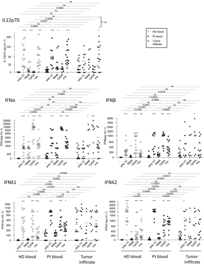
Figure 7.

Enhanced secretions of IL12p70, type I and III IFNs both with and without ex vivo TLR triggering arised from circulating and tumor‐infiltrating cells of melanoma patients. Cell suspensions from blood (HD, n = 18, open circles; Pt, n = 15, filled circles) or tumor infiltrates (Pt, n = 15, filled triangles) were stimulated for 20 h with or without TLR ligands (polyI:C, R848 or CpGA) alone or mixed together, and the culture supernatants were examined for the presence of IL‐12p70, IFNα, IFNβ, IFNλ1 and IFNλ2 by Luminex technology. Results are expressed in pg mL–1. Bars indicate mean. Stars indicate significant differences of the stimulated conditions compared to unstimulated ones within each group. P‐values were calculated using Mann–Whitney (dashed lines) and Kruskal–Wallis with post hoc Dunn’s multiple comparison (stars) nonparametric tests. *P ≤ 0.05, **P ≤ 0.01, ***P ≤ 0.001.
