Figure 5.
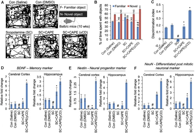
CAPE improves the cognitive behavior and memory function in scopolamine (SC)-treated mice models of neurodegenerative disease. (A) Readouts of novel object recognition (NOR) test showing the map of mice activities with familiar (F) and novel (N) objects in control (saline, DMSO), SC-treated, and CAPE-treated, CAPE-γCD-treated groups given treatments after SC exposure. CAPE post-treatment after SC exposure in mice was observed to improve cognitive behavior and memory function. (B) Quantitation showing percentage of time spent by mice with familiar and novel objects, while CAPE-γCD post-treatment found to improve mice’s attention to new objects. (C) Quantitation of object discriminative ability of mice showing improved ability with CAPE-γCD post-treatment in SC-treated mice. (D–F) Quantitative RT-PCR data showing expression of BDNF (D), Nestin (E), and NeuN (F) markers in the cerebral cortex and hippocampus regions in mice brain in control, SC, CAPE, and CAPE-γCD post-treated groups before SC treatments. Increased mRNA expression of these markers was observed in CAPE-γCD, and a bit lesser with CAPE post-treated groups after SC exposures. Histogram represents mean of the data (±SEM). Statistical analyses were performed using one-way ANOVA followed by post hoc student’s-Newman–Keuls test. #p < 0.05 and *p < 0.05—significant difference as compared with saline control and SC, respectively. The data were expressed as mean ± SD. The ##indicated p < 0.01 (familiar object as compared to SC), $$indicated p < 0.01 (novel object as compared to control-saline), and indicated ++p < 0.01 (novel object as compared to SC). NS indicated non-significant correlation.

