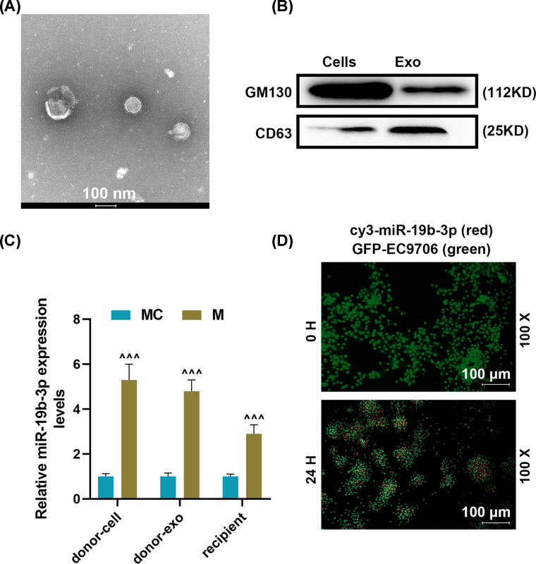
Figure 2. Effects of miR-19b-3p derived from exosomes on recipient human esophageal cancer cells (EC9706).
(A) Identification of exosomes by transmission electron microscope. (B) Western blot was used to detect surface markers of exosomes. (C) RT-qPCR was used to detect miR-19b-3p expression in mimic control (donor cell), miR-19b-3p (donor cell), mimic control (donor exo), miR-19b-3p (donor exo), Mimic control (recipient), miR-19b-3p (recipient) group. (D) Fluorescence microscope was used to observe the transmission of cy3-miR-19b-3p between EC9706 cells through exosomes. U6 was used as the internal reference gene of miRNA. Results were expressed as the mean ± SD; RT-qPCR, reverse transcription real-time quantitative polymerase chain reaction; ∧∧∧P<0.001 vs. MC; MC, mimic control, M, miR-19b-3p mimic.

